
LIGNES/auto is going to go (slightly) off the beaten track, but it’s for a good cause that closely affects its editor’s entourage, like so many other people. There are around 80 diseases resulting from a malfunction of the immune system. The immune system attacks the normal constituents of the body. Faced with these complex diseases, such as multiple sclerosis, researchers are developing new therapeutic strategies with the aim of controlling the immune system without preventing it from standing guard against pathogens. Among these ‘tools’, MRI remains one of the keys to progress in research. The Institut du Cerveau, below, based at the Pitié Salpêtrière hospital in Paris, is at the cutting edge of this field, as we will discover when we also talk about… Tesla. Even though we’re moving a long way from design, by the end of this post you’ll feel a lot smarter.
To find out more about the Institute and/or to make a donation, click here: https://institutducerveau-icm.org/fr/

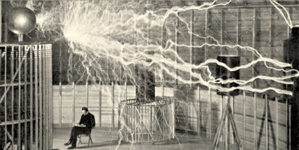
So I’m going to talk to you about MRI (many of you have undoubtedly sat in this tube at some point, which can sometimes be a cause for concern) and more specifically about… Tesla. Not Elon Musk’s Tesla, but that of the Tesla unit of measurement, named in honour of the Serbian physicist Nikola Tesla (1856-1943), famous for having put into practice the discovery of the wave nature of electromagnetism. This unit of measurement is the one used in the design, development and manufacture of the MRI core.
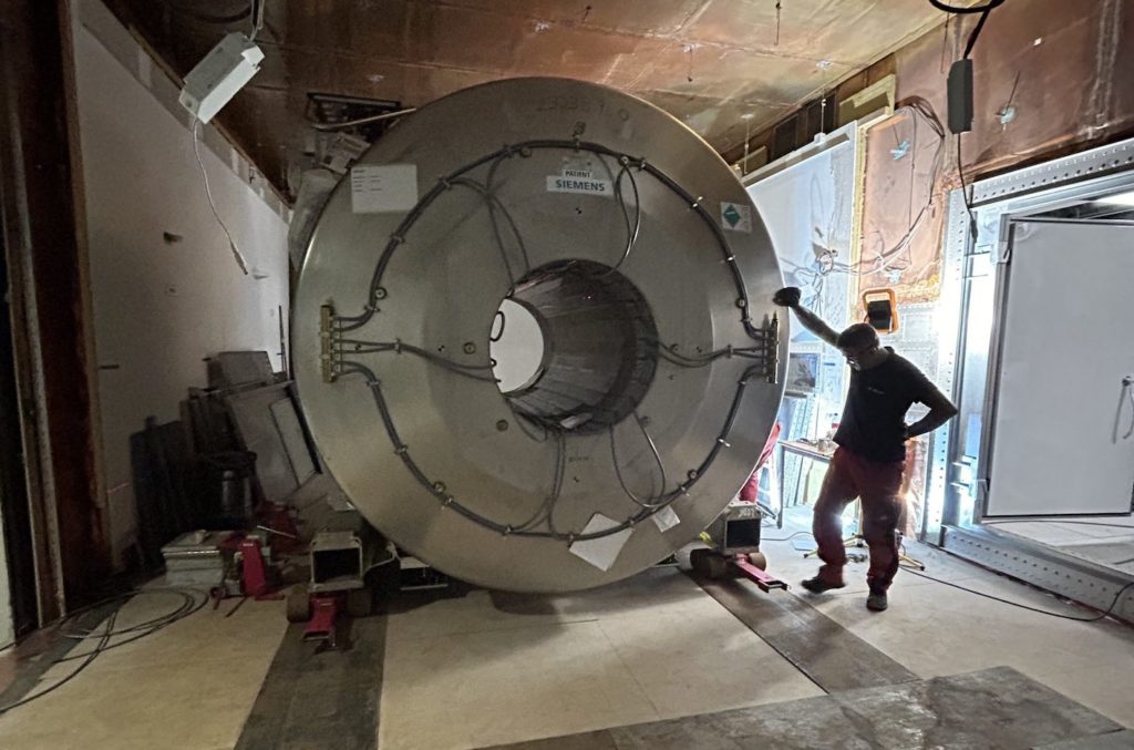
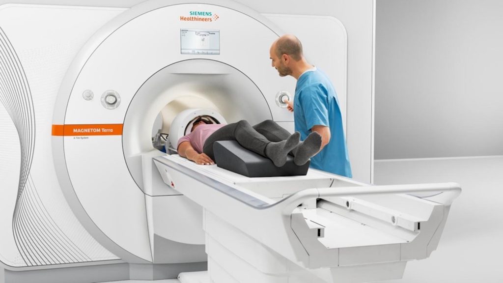
In France, the Institut du Cerveau in Paris received the very latest generation of MRI on 9 June: the 7T Magnetom Terra X MRI (Siemens Healthineers). It works with a magnet weighing around twenty tonnes (!) into which the patient slides! This magnet consists of a superconducting niobium-titanium alloy coil in a bath of liquid helium at very low temperature, creating a magnetic field equivalent to 140,000 times the Earth’s magnetic field… Hence the name 7T, T for… Tesla.
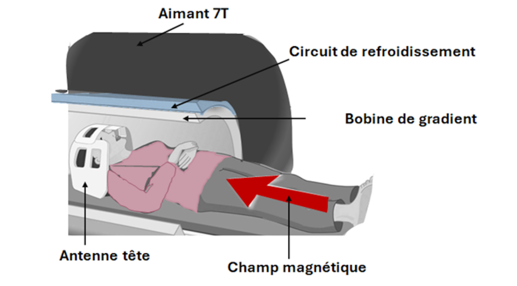
But if these figures are staggering, we first need to understand how they come about: how does an MRI scanner work? The Institute explains: “The magnet is at the heart of how an MRI scanner works. This magnet creates a magnetic field with an intensity of 7 Tesla. By comparison, the Earth’s magnetic field is only 0.00005 Tesla. Just as the Earth’s magnetic field orients the needle of a compass towards North, the MRI’s magnetic field, known as the main field, allows the hydrogen atoms contained in the water molecules of the body of the person being scanned to orient themselves along an axis parallel to the tunnel”.
“Electromagnetic waves called radiofrequencies, transverse to the main field, modify the magnetic properties of hydrogen atoms in a transient manner.The return to a state of equilibrium generates a magnetic signal that is recorded by an antenna.The way in which the atoms ‘respond’ to these waves and the intensity of the MRI signal generated depend on the properties of the tissue in which the atoms are located.The MRI signal is then recorded by a receiver coil and ‘translated’ by the machine into 2D or 3D images.The result is the detection – or not – of anomalies in the different areas examined, in this case the brain.
“Installing an MRI scanner imposes a number of constraints on its smooth operation.The main magnetic field must be strictly confined to the room in which the MRI is located, for safety reasons, so as not to attract or interfere with the operation of metallic or electronic components passing nearby. To achieve this, the Institute specifies that the walls and floor of the room have been covered with shielding made of an alloy called “mumetal”, composed of 20% iron and 74% nickel”.

In addition, in order to produce a quality signal that can be used for medical and scientific imaging, the MRI must be protected from all external electromagnetic interference by a Faraday cage made of copper foil. The control and acquisition room, where the radio manipulators operate the equipment, has a window overlooking the MRI room, fitted with glass that is hermetically sealed against electromagnetic fields.
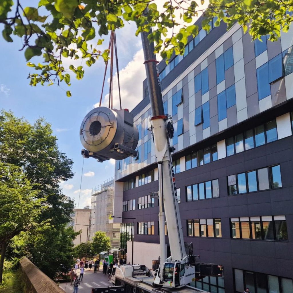
And installing the heart of this MRI, our famous magnet weighing around twenty tonnes, was no easy task in the middle of Paris! The Institute points out that it required “the dismantling of an exterior wall and interior partitions of the building to reach the MRI’s destination room”. But this baby is still one of the lightest in the world, as the Institute points out: “The 7T MRI magnet installed at the Brain Institute is the latest generation of this technology. It is the lightest magnet of this intensity in the world. It uses zero helium evaporation technology, thanks to a system that recondenses helium vapour into liquid helium.
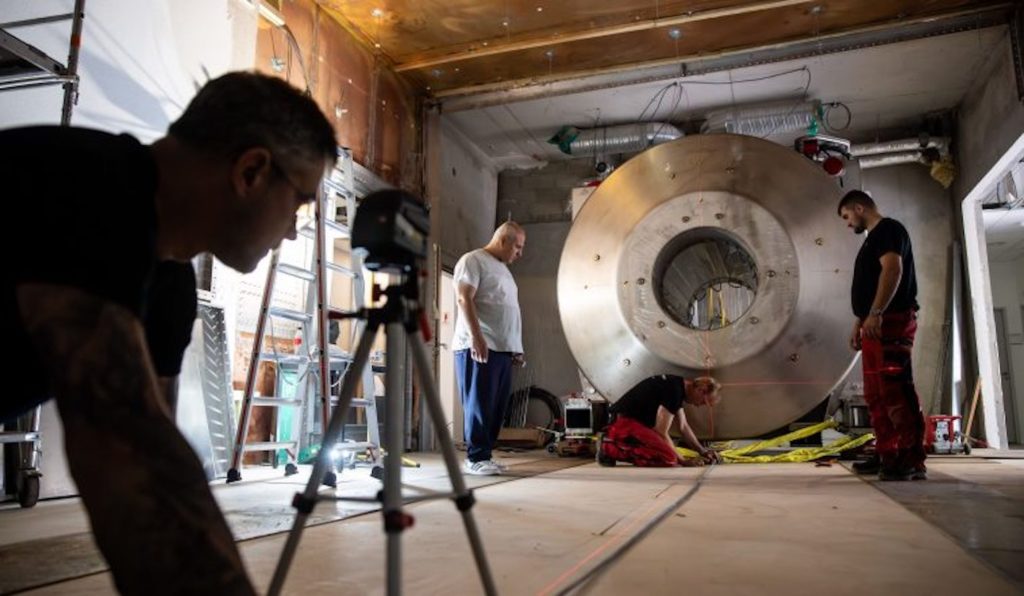
“Once the magnet’s field strength has been reached, scheduled for the summer of 2024, a phase of adjustment and fine-tuning of the device will be necessary, thanks to the expertise of the CENIR platform staff and an engineer from Siemens Healthineers seconded full time to the Institut du Cerveau. This adjustment phase will enable us to optimise the application of this technology to brain and spinal cord exploration”. Ultimately, the first research protocols should begin in early 2025. The expected technological leap lies mainly in the increase in the signal-to-noise ratio, enabling us to observe tissue structures smaller than a millimetre, thanks to the improved spatial resolution of 7T MRI, and to obtain images of molecules whose signal is too weak at lower fields, and in the improvement in image contrast, enabling us to image structures that are invisible at lower magnetic fields”.
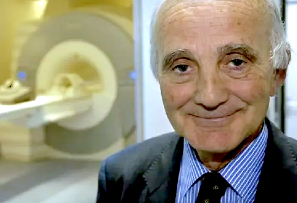
Finally, the Brain Institute was founded in 2010 by Gérard Saillant, Professor of Orthopaedic and Traumatological Surgery and now President of the Institute, and Jean Todt.Its founding members include other leading figures from the world of motor sport, such as Michael Schumacher and Max Mosley, and it has been supported by Alpine F1…
